




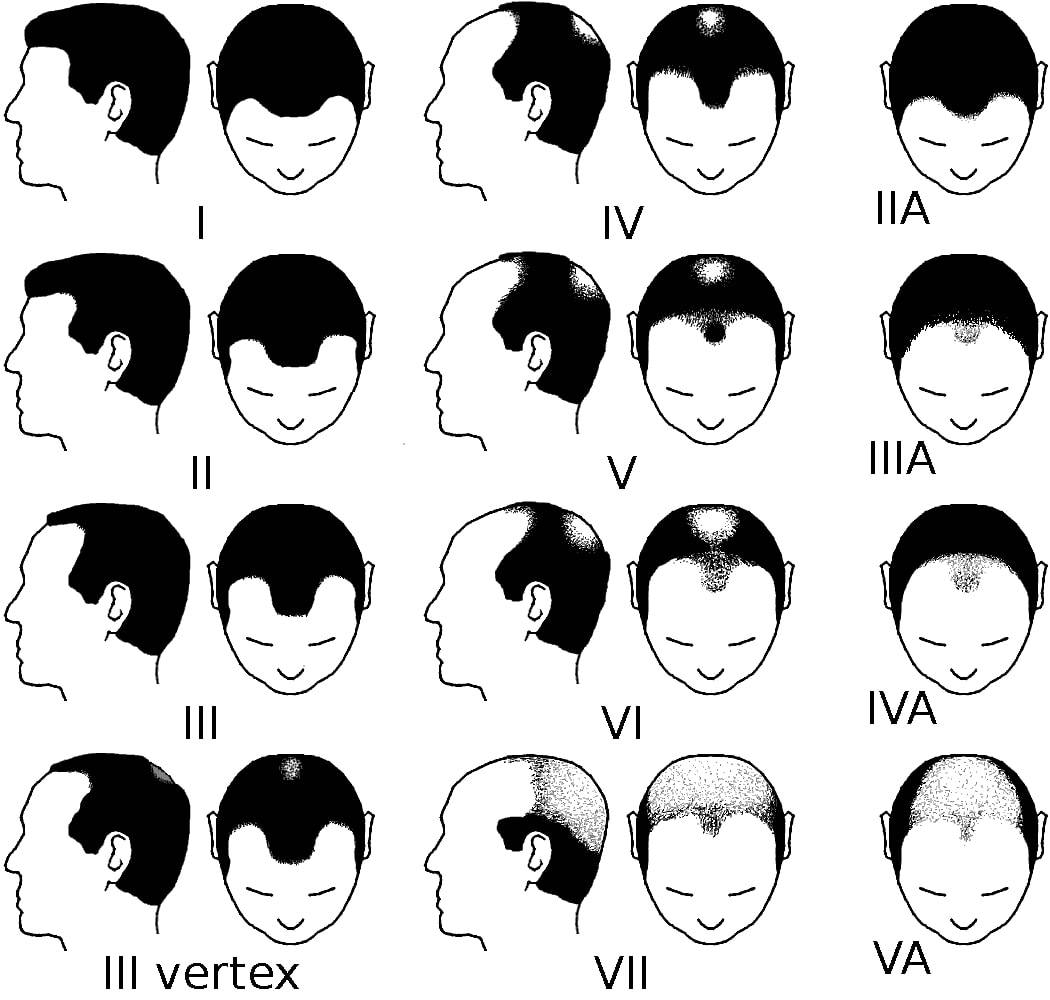
The Norwood Scale classifies the common pattern of male pattern hair loss in seven stages, from minimal temple recession through to extensive loss across the front, mid-scalp and crown. Hair transplant surgeons and clinicians use it as a reference point of describing what they can see. It also gives them an initial idea of what treatment is potentially going to be appropriate for the patient.
What it cannot do is explain why hair loss is occurring, confirm that androgenetic alopecia is the cause, or determine whether treatment is appropriate. A stage number describes a visible pattern. It does not measure donor density, predict how loss will progress, or define what surgery can achieve.
Two men at the same Norwood stage can differ substantially in age, hair calibre, donor density, rate of progression, family history and the amount of native hair still present between thinning areas. At Harley Street Hair Transplant Clinics, both Dr Elgindi and Dr Ditta assess all of these factors before any treatment discussion begins.
What is the Norwood Scale ?
The Hamilton–Norwood Scale classifies the recognised pattern of androgenetic alopecia in men. Two areas commonly change together: the hairline moves back at the temples, and the crown gradually becomes thinner. As loss advances, the frontal and crown regions widen and can eventually merge into a single continuous area.
The scale runs from Stage 1, where no recession is present, through to Stage 7, where only a narrow band of hair remains at the back and sides. The seven stages proceed as follows:
- Hairline intact. No recession at the temples or thinning at the crown.
- Slight recession at the temples often called a mature hairline.
- Deeper recession creating an M, V or U-shaped hairline. The first stage regarded as clearly established male pattern hair loss.
- More pronounced frontal recession and a larger crown area, with a band of native hair in the middle (sometimes called the bridge) still separating the two.
- The band between the frontal and crown areas (the bridge) has narrowed significantly or become less dense.
- The frontal and crown areas have merged. A continuous area of loss covers the front, top and crown.
- Only a horseshoe-shaped band of hair remains at the back and sides.
Clinicians use the scale to record what they observe and to discuss it in consistent terms. A stage number helps frame the conversation for the patient. It does not, by itself, determine what surgery can achieve or whether it is appropriate.
Why two men at the same stage can look very different
The Norwood stage describes the outline of hair loss, not its appearance. Hair calibre, colour contrast between scalp and hair, the density of remaining native hair and how that hair is styled all affect how much scalp is visible, even between two patients at the same stage. A man with fine, light-coloured hair may show more scalp at Stage 3 than a man with coarse, dark hair at Stage 4.
Photographs are also easy to misread: wet hair, unflattering lighting and camera angle can make loss appear more or less advanced than it is. This is why a scalp examination, under consistent conditions, remains the basis of any proper assessment.
Norwood Stage 1
The frontal hairline remains intact at Stage 1, with no visible recession at the temples and no thinning at the crown. This is the baseline the classification uses. It does not mean that hair loss will or will not develop.
If somebody at this stage notices increased shedding or a change in density despite an unchanged hairline, the Norwood Scale may not fully describe what is happening. Diffuse thinning and sudden shedding are assessed separately. At Stage 1, there is usually no surgical issue to address. Consistent photographs taken in the same lighting and angle can provide a useful baseline if the hairline appears to change later.
Where hair loss is a concern, a clinician may discuss medication. Finasteride works by reducing the conversion of testosterone to dihydrotestosterone (DHT), the hormone associated with follicle miniaturisation. Minoxidil acts differently as it is a hair growth stimulant, not a DHT blocker and the two are often used together. Neither is appropriate for every patient and both require an ongoing commitment to maintain any benefit.
Norwood Stage 2
Early temple recession
The hairline has moved back slightly at the temples, often in a broadly symmetrical pattern. This is sometimes described as a mature hairline. The term does not, on its own, indicate whether recession has stopped or is continuing.
A single photograph cannot show whether the hairline is stable. Comparing images over time works best when hair length, camera angle and lighting are kept consistent. A clinician will also ask when the change began and whether it appears to be progressing. Surgery is rarely discussed at this stage, and medication, where appropriate, remains the most common course of action.
Planning a low, dense transplant this early carries risk. If native hair behind the transplanted line later becomes thinner, the result can look unnatural as the pattern advances. Any surgical discussion must account for the likely long-term pattern, not only the present hairline position.
Norwood Stage 3
The third stage on the Norwood scale is what can typically be defined as early male pattern hair loss. This is where further hair loss takes place beyond the “adult” or “mature” hairline as noted in stage two.
At this stage, your hairline will recede further up the temples causing a frontal tuft at the front of the head. Generally speaking, patients also feel that their hair begins to thin or feel finer to the touch.
At this stage, surgeons may begin to recommend a hair transplant as an option. However, this will dependent upon your family history, as this is the best indicator as to how your pattern hair loss is most likely to progress. Although, a hair transplant is a long-term solution to pattern baldness, it is important to consider how your hair will look in the future.
Stage 3 (Vertex)
Crown thinning alongside frontal recession.
Stage 3 Vertex includes a defined area of thinning at the crown alongside the frontal changes already present at Stage 3. The crown follows a natural spiral growth pattern and its apparent size can change considerably depending on lighting, hair length and styling, making it one of the more difficult areas to assess from photographs alone.
Crown loss can spread across a large circular area over time. For younger men, treating the crown too early carries a specific risk: as pattern hair loss continues beyond the transplanted zone, a ring of thinning native hair can appear around the restored area (a visible gap between the transplant and the surrounding scalp, sometimes called the “halo” effect) that becomes harder to correct. This is why crown hair transplants need long term planning.
The front and crown also draw from the same finite donor supply. Committing grafts to the crown at this stage may reduce what is available to maintain the frontal frame later, a decision that has to be made with the whole scalp pattern in mind, not the crown in isolation. Medication is typically the first recommendation for younger men experiencing early crown thinning.
Norwood Stage 4
Separate frontal and crown areas.
Recession at the front is more pronounced and the crown has a larger area of loss. A band of native hair still separates the two areas across the mid-scalp, but its density needs to be examined rather than assumed. Hair that appears full when combed forward may be considerably thinner underneath.
At this stage, priorities have to be agreed clearly before any surgical plan is made. The size of the recipient area is growing and the donor supply is fixed. Rebuilding the frontal frame tends to have more visual impact than spreading the same number of grafts across the hairline, mid-scalp and crown simultaneously.
The patient’s aims must remain within what the donor area can realistically support. Patients reaching Stage 4 should generally plan for the likelihood that loss will continue to progress, since Stage 4 is often the beginning of a longer trajectory towards Stages 5, 6 and 7.
Norwood Stage 5
A narrower bridge of hair.
The frontal and crown areas have grown larger and the band of native hair between them has become noticeably narrower or less dense. The two areas have not yet fully joined, but the surface requiring coverage is now substantial.
At Stage 5, the balance between coverage and density becomes a central part of any surgical discussion. Attempting to restore the density of a full head of hair across the entire bald area is rarely an appropriate use of the donor supply. A surgeon needs to identify where grafts will make the greatest visible difference (usually the frontal area) and where lighter coverage may be a more sustainable outcome.
Donor measurements matter more than a promised graft count at this stage. The result that can be achieved depends on actual density, hair calibre, colour contrast between hair and scalp, and safe extraction limits ,not a number quoted before a proper donor assessment.
Stage 6
The front and crown have joined.
The strip of native hair across the mid-scalp (bridge) has largely disappeared, leaving one continuous area of loss covering the front, top and crown. Hair remains around the back and sides of the scalp. This is the permanent donor zone.
The scale of the recipient area makes prioritisation unavoidable. Concentrating grafts in the frontal third frames the face and provides the most visible restoration. The mid-scalp and crown may require a lighter approach, accepting less density in exchange for broader coverage. Full native-looking density across the entire bald area is not a realistic outcome at Stage 6, and any surgical plan should be agreed on that basis.
Surgery remains possible at Stage 6 (at least a 2 stage surgery approach) when the donor area is strong, but it requires a conservative hairline position and clearly agreed expectations. The aim is strategic coverage, not complete restoration.
Stage 7
The most advanced pattern.
A narrower horseshoe-shaped band of hair remains at the back and sides, and the donor fringe may sit lower on the scalp than it does at Stage 6. The gap between the size of the recipient area and the available donor supply is at its greatest.
The donor area must be examined carefully for density and miniaturisation. Follicles at the edges of the safe donor zone may themselves be susceptible to DHT meaning extraction from those areas carries the risk of depleting the back and sides without providing lasting coverage on top.
Most (if not all) patients at Stage 7 will not be suitable candidates for surgery as at this stage just the pure graft requirements exceed what can safely be harvested from the donor area by around half. So at stage 7, a patient will require between 12,000 and 14,000+ grafts for complete coverage. We can safely extract between 6,000 and 7,000 grafts from the scalp donor area before donor thinning becomes too apparent.
How the Norwood Scale is used in treatment planning
The scale helps a clinician record the present pattern of loss and discuss what may happen if androgenetic alopecia continues to progress. It also makes visible the central limitation of hair transplantation: the donor area is finite, but the area that might eventually require coverage can continue to expand.
A surgical plan therefore considers far more than the visible bald area. Donor density, evidence of miniaturisation, hair calibre, scalp laxity, family history, age, medical history, any previous surgery and the patient’s own priorities all affect what can be planned. The Norwood stage gives the conversation a starting point. It does not determine the outcome.
What the Norwood Scale cannot tell you
The scale cannot confirm that male pattern hair loss is the cause of what you are seeing. Sudden shedding, patchy loss, scarring alopecia, scalp inflammation or an unusual distribution pattern all require a broader clinical assessment that goes beyond Norwood staging.
The classification also does not measure donor strength, predict the exact graft requirement for a given result, or establish whether somebody is suitable for surgery. It was developed around the typical male pattern hair loss and diffuse thinning across the scalp may not fit neatly into any Norwood stage. Female pattern hair loss is usually classified differently.
Photographs and self-assessment against stage diagrams are a reasonable starting point, but they are easy to misread. Wet hair, lighting conditions, camera angle and hair length all change the apparent density and extent of recession. A proper assessment involves a scalp examination with a trichoscope, a donor area evaluation and a clinical conversation, not a stage number alone.
Summary
The Norwood Scale is a useful starting point, a common language that lets clinicians and patients discuss visible hair loss clearly. What it cannot do is substitute for a proper assessment. A stage number tells you where you are. It does not tell you where you will be, how much donor hair you have available, or what any surgical procedure can realistically achieve.
Hair transplant planning is fundamentally a question of surface areas: the size of the area requiring coverage relative to the density and quality of the donor zone. A larger recipient area does not automatically mean surgery is impossible, but it does mean that priorities have to be set and that the expectations agreed at the outset need to reflect what the donor supply can genuinely support.
If you would like to understand where you sit on the Norwood Scale and what the options are for your specific pattern of loss, get in touch with us for an initial consultation with one of our patient advisers and subsequently, one of our hair transplant surgeons. An assessment covers the full picture, not just the stage.